Is a nap as good as a night?

Mednick, S., Nakayama, K., & Stickgold, R. (2003). Sleep-dependent learning: a nap is as good as a night. Nature neuroscience, 6(7), 697.


Review

Is getting enough sleep really that important? According to the CDC, 1 and 3 Americans are not sleeping as much as they should be every night. For quite some time, the function of sleep has eluded us. This, perhaps, has led us to disregard its benefits and continue with a life of sleep deprivation. However, in the past 20 years, research has made astounding progress in understanding what exactly sleep does for our body and mind. Recent findings especially have highlighted sleep’s importance and what we can do to circumvent the sleepless side effects of the typical busy American life.

A study published back in 2003 by Dr. Sara Mednick entitled, Sleep-dependent learning: a nap is as good as a night, examines one particular aspect of sleep known as memory consolidation. Research has shown that sleep not only repairs our body but also replenishes the mind though a serious of neural processes that keep us sharp and alert during the day. Memory consolidation is but one of these many neural functions, and operates by solidifying daily acquired skills and memories into long-term storage. You can think of this process as analogous to storing files on your computer’s hard drive or installing new software that requires a reboot after completion. Similarly, the human brain also requires a “reboot” to utilize new learning to its maximum efficiency.


Visual learning and sleep

Dr. Mednick and colleagues employed their investigation of sleep dependent memory consolidation in the field of “perceptual learning,” a variation in vision science that focuses on how our visual system learns new information in the environment. Perceptual learning has been widely studied for decades and acts as a powerful tool in testing brain plasticity due to how well we now understand the visual system and its functions. In the present study, Mednick tested over 70 healthy participants on a standard perceptual learning task, referred to as a Texture Discrimination Task (TDT), and assessed how naps affected performance trends.

The experiment consisted of three groups. The first group was asked to arrive at the Harvard sleep lab at 9:00am, where they were then asked to complete roughly an hour of training on the TDT. Their data over that hour was collected and fitted to a psychometric function allowing the researchers to determine a “threshold” in performance, which specified the hardest difficulty level on the TDT that the participants could still keep 80% accuracy. The first group of participants was then asked to come back to the lab at 7:00pm that evening to complete a short “re-test” to measure performance changes and then again that following morning at 9am. The schedule for the second and third groups was identical to the first, except that they were asked to come back to the lab at 2pm to take a nap for either 60 minutes or 90 minutes, respectively. While seeping, the researchers monitored the participants’ brainwaves on a monitor with an electroencephalogram (EEG). In sum, the experiment investigated TDT performance changes over a 24-hour period with a no-sleep control group, as well as with 60-minute and 90-minute nap experimental groups.


Stages of sleep

One of the major benefits of this design plays on the fact that sleep is naturally broken up into various stages over an average of 90 minutes. While the exact values are different for everyone, your body cycles between four stages every 90 minutes that occur roughly 5 times over a typical night’s sleep. In each cycle, the body first enters light sleep known as Stage 1 sleep, where it is easiest to be disturbed and woken up. Stage 2 sleep then follows, and marks the beginning of deeper sleep, lasting approximately 20-30 minutes in duration. After, the body enters Stage 3 sleep, known as Slow Wave Sleep (SWS), which is marked by giant, synchronous delta brainwaves that can be seen on an EEG. SWS is considered to be the deepest part of sleep, even though the following stage, known as Rapid Eye Movement (REM) sleep, is thought to carry extreme significance for memory consolidation functions. By examining perceptual learning in the presence of naps, Mednick and colleagues were able to parse out subgroups that showed just SWS or both SWS and REM within the napping participants.

Screen Shot 2017-09-01 at 11.58.40 AM

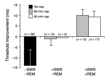
The results of Mednick’s study were quite novel, revealing unique trends in TDT performance associated with various sleep stages. The research team found that the groups that were able to experience both SWS and REM sleep during their naps had the largest improvement on the TDT. Interestingly, these same participants displayed even greater improvement when tested the following morning, far out-classing their SWS-only and non-nap counterparts. The SWS-only groups showed consistent performance on the TDT when re-tested at 7:00pm and only showed improvement the following day. This is similar to the no-nap group, except they actually performed worse on their 7:00pm re-test compared to the initial training, suggesting that the visual skills acquired were deteriorating without the memory-solidifying presence of sleep.

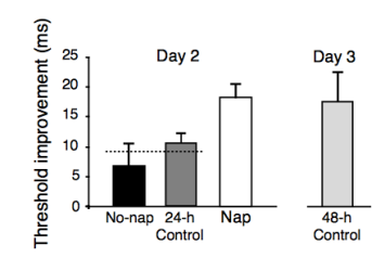
Control groups were employed both 24 hours and 48 hours after the initial training on the TDT, showing similar tends to the group that was able to achieve REM sleep in addition to SWS during their nap. The research team concluded that one full nap containing both SWS and REM sleep was sufficient to cause plastic changes in the brain that allowed for improvement on the TDT. This improvement was not only significant, but was almost identical in magnitude to the performance changes seen over 24 and 48 hours. This magnitude of improvement was unforeseen, and opens up many possibilities for the benefits of regular naps.

Screen Shot 2017-09-01 at 12.48.09 PM


Are naps all we need?

The present study seems to heavily suggest, as seen in the title, that naps are indeed as good as a night. While this is quite a stretch from the truth in the literal sense, the researchers at Harvard University demonstrated that one full cycle of sleep is highly beneficial to acquiring visual skills. They speculated that there is something unique to REM sleep that affects the consolidation process of TDT. One theory is, given that TDT is a “procedural skill,” which means that is in the same realm as learning to play an instrument or ride a bike, that REM sleep specifically deals with procedural memories and their subsequent consolidation. Other studies have found that certain stages of sleep may be specific to other kinds of skills and memories, such as explicit memory, where memorizing lists of information is the core goal.

One major complication with this research is the multitude of studies that seem to support opposing findings depending on the task or experimental design. Naps with perceptual learning has been covered in a limited fashion, but analogous learning experiments have been conducted using different modalities such as motor learning, which has been regarded in classical research as similar in characteristics to perceptual learning. These studies (conducted by Dr. Giulio Tononi and colleagues) illustrated a plethora of evidence that SWS is actually the key to performance improvement on various tasks. Other groups have reported that, rather than each individual stage, the relationship between SWS and REM sleep is what allows for successful consolidation.

Mednick’s study alone, does not resolve any of the above controversies, but instead offers a new perspective on studying sleep with their unique nap design. Should we substitute our debilitating daily fatigue with a nap rather than a full night of sleep? Probably not, but what we can take away is that a nap, especially one with a full sleep cycle, is never a bad idea.

Inception: fantasy or fact?

Shibata, K., Watanabe, T., Sasaki, Y., & Kawato, M. (2011). Perceptual learning incepted by decoded fMRI neurofeedback without stimulus presentation. science, 334(6061), 1413-1415.


Review

The word “inception” probably makes you think of Leonardo DiCaprio’s 2010 dream-weaved psychological thriller, where a technique for “planting information” in the brain is revealed and explored. After the release of Leo’s movie, few would have predicted that the word “inception” would be found in factual scientific literature only a year later. In the present study entitled, “Perceptual learning incepted by decoded fMRI neurofeedback without stimulus presentation” authored by Dr. Takeo Watanabe, we find that inception is no longer a word only found in fantasy, but exists as tool in frontier neuroscience.

Dr. Watanabe’s “inception” involves a specific branch of neuroscience known as the study of “visual perceptual learning” or “VPL” for short. VPL is, in the general sense, how our visual system uses new information to adapt and become maximally efficient given the constraints of the environment. The present study utilizes the science of VPL through an extraordinarily clever design that even challenges the famed “correlation is not causation” debate in one swift experiment.

In the typical fashion of research, it is extremely important to pick and choose the tools with which to illustrate scientific data that best conveys the broader scope of the project. In this case, Watanabe and colleagues used little grey and white distortions called, “Gabor patches” as a measure of how well they were able to incept their participants. These visually noisy, patchy gratings have been used in the field of VPL for over 20 years and are believed to engage one of the most basic functions of the visual cortex: orientation detection.


Inception from the bottom up

The visual cortex is built from the bottom up. The most basic features are recognized by parts of the early visual cortex whereas higher areas process more complicated features such as color and motion. Watanabe’s team chose the early visual cortex for their experiment due to the controversial nature of whether or not this part of the brain can still learn in adulthood, since it was once believed that only certain parts of the brain were plastic after early development.

Demonstrating learning through the early visual cortex would not only provide invaluable evidence for this controversy, but would also suggest tendencies for plastic nature throughout the rest of the brain.

On the first day of the experiment, deemed the “pre-test”, wide-eyed and unknowing participants were shown three types of patchy gratings (known in colloquial research as “Gabor patches”) with 10, 70, or 130 degree oriented lines. These three orientations, upon viewing, engage specific cell lines in the early visual cortex than can be detected through brain imaging techniques. During the pre-test, participants were asked to report which of these three orientations they saw as the patches were progressively drowned out with visual noise to the point where the orientation was almost invisible. Accuracy was based on how well the participants could correctly discriminate the orientations at high noise levels. This first day of experimentation was identical to the last day (the “post-test”) allowing the researchers to assess how much the participants improved over the course of the entire experiment.

incept1.png

Construction

The second day of Dr. Watanabe’s experiment marked the beginning of a scientific process most would consider fictional and quite frankly, impossible. Participants were asked to lie in an MRI scanner while the researchers recorded brain activity from their visual cortices. The research team however, was not searching for just any activity, but instead equipped the scanner with a video screen and presented our three familiar Gabor patches to the participants. As each orientation passed in front of a participant’s eyes, the MRI outputted a unique pattern that was derived from brain activity in the early visual cortex in its attempt to recognize each orientation. The researchers were left with three distinct brain activity patterns that represented internal processing of each Gabor patch.


Induction

Now that the data was acquired, the fun could begin. The participants underwent 5 to 10 days of training depending on their designated group (4 participants conducted 5 days and 6 conducted 10 days of training) without the knowledge of what the researchers were attempting to accomplish. During each day of training, the participants would return to their cozy MRI tube and were presented with a video display containing nothing but a round green disk. They were then given an instructionally vague task, where the experimenters directed them over a loudspeaker to, “somehow regulate activity in the posterior part of the brain to make the solid green disc that was presented 6 s later as large as possible.”

As difficult as the request may have seemed, the researchers made sure to sufficiently motivate their participants by including a bonus payment proportional to the average size of the disk during the experiment. Sure enough, each participant learned how to focus his or her thoughts to enlarge the green disk on the video display over several days of training in the MRI scanner.

As mentioned above, the last day of the experiment included the Gabor orientation test that was conducted on the first day. The participants found themselves particularly adept at discriminating one of the three orientations, differing greatly from performance trends collected at the beginning of the experiment.

The reasoning behind these results can be found in the making of the fabled green disk. The researchers used a pattern classifier to take the average brain activity from viewing one particular Gabor patch in the induction phase and then compared it to real-time data from the scanner during training. The more similar brain activity was to the induction phase during the training, the larger the size of the green disk. In short, the size of the green disk represented the brain activity that occurs when viewing a particular Gabor patch.


Inception in modern science

The research team, under Dr. Watanabe’s directive, was able to incept visual training into an unknowing participant’s mind so that they actually became better at a task without even knowing they were training for it.

Not only were the participants unaware of what orientation they were given, but they reported using strategies during training that differed greatly from the content in pre-test. Some participants noted they thought about traffic lights, some reported visualizing scenery. Many included mental imagery that bared little resemblance to any Gabor patch features. It seemed that as long as that particular area in the visual cortex corresponding to Gabor orientation was activated, then learning occurred.

The results from this study demonstrated several key findings that challenged years of hotly debated scientific content in one cleverly designed experiment. One of the largest controversies in scientific data interpretation, known as the issue of “correlation is not causation,” was effectively eliminated as a possibility from this design. Studies using single-unit recording techniques demonstrated correlation between brain activity and behavior, whereas trans-magnetic cranial stimulation (TMS) experiments have shown that a particular brain region is necessary for a behavioral by temporarily inhibiting its function. Inception, however, effectively shows causation by creating a new behavior after a particular brain region is engaged for a certain amount of time, even without the participant’s knowledge of the task.

The use of inception in Dr. Watanabe’s study has demonstrated adult visual plasticity, a casual role for visual learning, and opened up a wide range of new possibilities for adapted use. Particularly, scientists could “incept” a person with new skills, memories, or even rescue cortical functions non-invasively. The future could hold a number of uses for this new technique, bridging the gap between fantasy science and real world application